waynakyo
Experienced Member
- Reaction score
- 465
Digital microscopes are cheap these days. So I got me one since i am thinking about hair transplant.
But I found yellow dots in the thinning area. And apparently there is a recent study on that:
I wonder whether the causation goes from hair thinning => engulfed sebaceous glands (as the paper seems to be suggesting) or it is the other way.
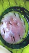
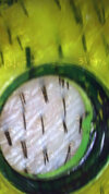
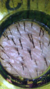
I attached a picture of the healthy donor area (no dots) and two of the thinning area. I need to shave the latter for better view.
But I found yellow dots in the thinning area. And apparently there is a recent study on that:
Yellow dots in trichoscopy: https://www.ncbi.nlm.nih.gov/pmc/articles/PMC5674712/
I wonder whether the causation goes from hair thinning => engulfed sebaceous glands (as the paper seems to be suggesting) or it is the other way.
I attached a picture of the healthy donor area (no dots) and two of the thinning area. I need to shave the latter for better view.
Last edited:


